How can our research help to improve the diagnosis of osteoporosis fractures?
20 October 2020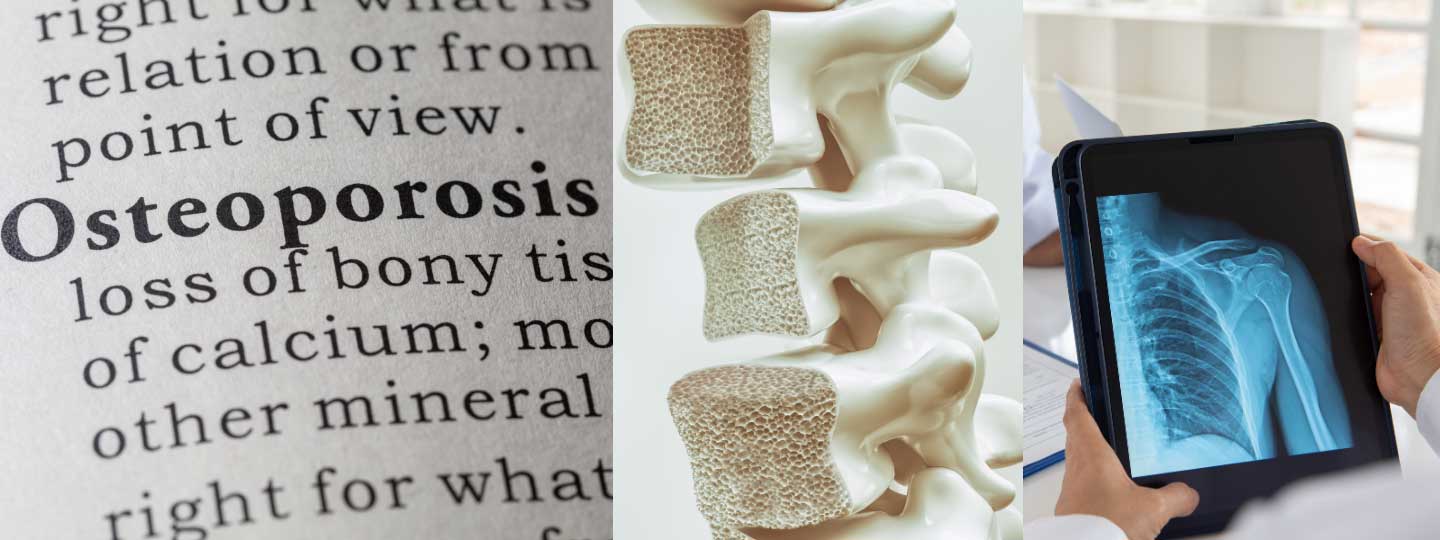
We wanted to celebrate one of our amazing research projects aiming to improve the lives of people living with osteoporosis.
It was hard to choose just one, but after searching through our extensive research, we decided to share the project taking place at the University of Leeds, led by Professor Alejandro Frangi, called the VERDICT study.
Why is this research needed?
Osteoporosis is common in the UK, and the risk increases with age. Anyone can get osteoporosis, but women are about four times more likely than men to develop it.
Osteoporosis leads to more porous bones, meaning the holes in the usual honeycomb structure within our bones become larger, and the overall density is lower. This means that the bones become more fragile and are therefore more likely to fracture or break.
A common fracture in patients with osteoporosis is in the spine, however, these fractures may sometimes go undetected as patients don’t suffer any symptoms.
Early detection is essential as once somebody has a first fracture, a second is more likely. Therefore, a tool is needed to detect these fractures, making sure they don’t go undiagnosed, and that the patient can receive the correct treatment quickly.
“We estimate that at least 5% of older people have vertebral fractures and yet don’t know about it. So, if the expertise were available using our AI software throughout the country, then we could diagnose fractures early and in many more patients.”
How will early fracture detection help patients?
Professor Alejandro Frangi is hoping to provide a solution to this problem through his research that we have funded, as there is currently no standard method to look at spinal fractures in people with osteoporosis.
Usually x-ray or dual x-ray absorptiometry (DXA) is used. DXA can provide measurements of bone mineral density, as well as showing the bone structure.
However, both these techniques are two-dimensional, and require specialist training. Therefore, mild fractures are sometimes going undiagnosed.
This research will develop a new computer aided approach, which will be three-dimensional, and will also be more user friendly, allowing small fractures to be identified.
Being able to diagnose spinal fractures early and consistently will mean that osteoporosis patients can receive the correct treatment quickly, and ensure their condition is monitored appropriately to prevent other spinal fractures from occurring.
What have they found so far?
The team assessed 500 people for osteoporosis and possible fractures, using the standard approach in which a radiographer or trained specialist assesses the two-dimensional scans.
The researchers then developed a computer-based approach capable of generating a 3D reconstruction of the vertebrae and analysed the spinal images in the same way.
Researchers studied the normal variation in the anatomy of the spine and trained the computer-based technique to account for these differences when identifying fractures. This helps to ensure that the new technique does not mistake normal differences in spine shape for a fracture.
The researchers have shown that their technique can identify fractures that are hard to detect or sometimes missed by the trained specialist.
Using this computer approach could help future treatments as it as it allows earlier detection of fractures and it would also free up time of specialist usually analysing these images.
Find out more about our research
We’ll keep you posted on progress and updates. In the meantime, find out more about our research.
Get the support you need
- If you would like to talk to someone, you can call our free helpline on 0800 5200 520 (Monday to Friday, 9am to 8pm)
- Talk to our arthritis virtual assistant, 24/7
- Join our online community
- Stay in touch and follow us on Twitter, Facebook and Instagram.